Summary
Anatomy of the legs
Anatomy all aspects of the body is a significant barrier for athletes, however it is crucial for both achieving physical equilibrium and aesthetic appeal. Understanding the anatomical structure of the major muscles is vital, and the lower extremities, including the legs, are an integral component of this knowledge. From an anatomical perspective, the term “they” only pertains to a certain region of the lower extremities extending from the knee to the feet.
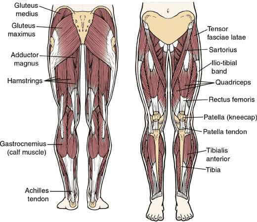
However, the lower extremities, which span from the hip to the feet, consist of many muscles such as the gluteal muscles, quadriceps, adductor muscles, hamstring muscles, calf muscles, and the iliopsoas muscle. These muscular structures function in coordination to uphold the body and facilitate locomotion, however their specific roles may differ according on the particular activity being performed, such as running, walking, or descending. This article will examine the components of the lower extremities in their conventional understanding.
The muscles of the thighs
Thigh muscle tissue When it comes to mobility and growth, the functions of these muscles are complementary to those of the buttock muscles, with which they are intimately associated or perhaps virtually inseparable.
To begin, the glutes are a set of huge, spherical muscles found near the lower back. Commonly known as the back, the anatomical term for this area is the gluteus maximus or gluteal region.
The huge gluteus maximus is easily identifiable; its size may be superficial, but its contribution to the gluteus’s curved form and overall strength is tremendous. In addition, it props up the thighs so the body is balanced.
The gluteal medium, which sits just below the gluteus maximus and secures the connection between the back and pelvic muscles, is another distinctive feature due to its fan-shaped morphology.
The thighs are another area it helps to stabilize. Although the deep-lying gluteus minimus contributes just a modest amount of total strength, it is crucial to abduction and rotation of the thighs. This group of muscles forms the hip joint by stabilizing the thigh bone’s (femur) connection to the pelvic skeleton.
Their indispensability is explained by their contribution to the body in maintaining balance, but also to the rotating motions of the hip. In addition, the muscles that support them supply additional power to the legs.
The quadriceps are a group of four muscular bundles located in the front of the thigh. It functions in the connection of the femur, patella, and tibia. Due to their crucial role in bearing the body’s weight, the hamstrings are often regarded as the strongest muscle in the human body.
The bundles that constitute it are the rectus femoris, vastus intermedius which is positioned in a deep plane and vastus medialis itself consisting of two lateral bundles. These systems help to keep the knees from buckling under too much weight and strain when standing.
The quadriceps are the backbone of the human body, making them crucial to the practice of any sport.
Another set of thigh muscles called the hamstrings. It covers an area that starts at the rear of the pelvis and goes all the way down to the lower legs. It acts in opposition to its paired muscle, the quadriceps, because it facilitates a flexing motion of the leg while the latter guarantees an extension.
In terms of their involvement in motions, the semitendinosus, semimembranosus, femoral biceps, or thigh biceps, limit full extension of the knee, slow down motion, and permit external rotation of the knee.
The adductors are a group of muscles located further inside the thighs. A set of muscles on the upper inner thigh that creates the characteristic curvature. There are three distinct muscle groups that make up the adductor magnus. The adductor magnus itself consists of tiny bundles deep within the thigh and is the largest muscle in the inner thigh in terms of surface area.
Next comes the superficial adductor magnus, and finally the adductor brevis, which consists of two bundles. It should be emphasized here that their existence guarantees adduction, rotation and flexion of the thigh.
In addition to their roles in flexion and internal rotation of the knee and leg on the thigh, these two muscles play a crucial role in adduction of the hip, thigh, and pelvis. When comparing the pectineus to the rectus internus, it’s important to note that the pectineus is a superficial muscle that runs from the iliac bone to the femur.
Pelvic muscles
The muscular networks in the leg area are very intricate, and certain muscles have an impact that reaches from the base of the spine to the groin. Its network enters the abdominal region and continues into the thorax.
The psoas major and the iliac muscle, which originate in the iliac fossa and terminate in the femoral portion of the thigh, combine to create the psoas-iliac muscle complex. However, there is a third muscle that plays a complementary, if smaller, function to the first two. The presence of this muscle varies from person to person, particularly if the latter has a well-muscled, dry abdomen.
Because it permits the bending of the hip and trunk on both sides, this muscle is significant.
Lower part muscles
The muscles that are most often neglected by many bodybuilders are the calves. Nevertheless, they play an equally important part in the dynamic movement. Although working on calf strength is simple and can be done anywhere and at any time without the need for any special equipment, stretching is an essential part of preventing tendinitis. They are sometimes referred to as the sural triceps, and their formation is the consequence of the assembling of three muscular bundles.
In terms of their anatomical composition, the bundles are made up of the twin muscles that are positioned on the surface, the soleus, which is located more profoundly, and the Achilles tendon, which is located inferiorly. These contribute to the substantial amount that it contains. These muscles are involved in many different forms of movement, most notably walking. For example, standing, flexing and elevating the ankle and heel, and pushing the foot forward while leaping are all examples of the roles that these muscles play in movement.
But they also play a role in guaranteeing the flexion and rotation of the knees, with the help of other series of deep muscles, such as the popliteus and the tibialis posterior, which are responsible for ensuring the flexions of the foot and the long flexor of the hallux.
READ MORE:
