Summary
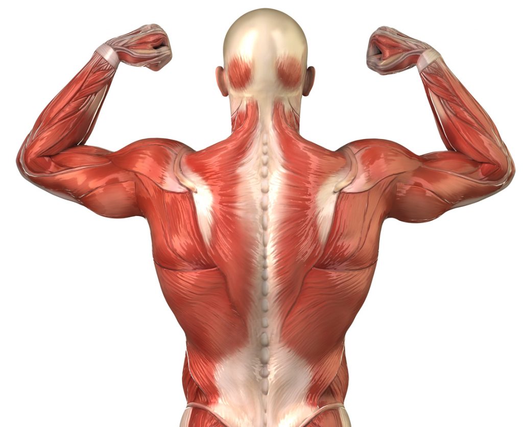
Anatomy of The Back
Anatomy of numerous muscle groups comprise the back. The massive dorsal muscles, trapezius muscles, and lumbar muscles are considered fundamental. The primary difference between each is its shape. For instance, the purpose of the massive dorsal muscles is to increase in size. The trapezius muscles comprise the uppermost region of the back. They consist of three fascicles specifically:
- The lower bundle
- the intermediate fascicle,
- the upper fascicle
The lumbar muscles are located in the lower back, opposite the trapezius muscles.
The lower back bone
The lumbar spine is the greatest muscle in the human body since it encompasses the entire back.
Aesthetically speaking, the lats is a significant muscle. In fact, the back’s V-shaped aspect is owed to it. Having strong latissimus dorsi muscles also aids in maintaining proper posture. Similar to the pectoralis major, this muscle joins the trunk to the forearm. Additionally, it joins the lower torso to the pelvis and the upper torso to the spine through a broad, flat mass of tendon known as the lumbosacral fascia.
The latissimus dorsi is a muscle that straightens the chest, which is another thing to keep in mind. The shoulders are drawn back and raised by it.
At the kidneys, the Latissimus dorsi ends in a fibrous blade. It is an auxiliary inspiratory muscle that enables the raising of the arms. The Latissimus dorsi is attributed to this merely because it is situated on the inside of the final four ribs. The bottom digits of the oblique abdominal muscles are also present.
It will also be seen when you do specific movements: this muscle extends the dorsolumbar spine and draws the shoulder blades together as you lower and raise your shoulders.
It is also known that the Latissimus dorsi muscle lowers the arm. In fact, without this muscle, your arm will naturally fall back down when you raise it. The Latissimus dorsi muscle helps to promote an extension movement when you lower your arm.
The muscle the trapezius
Placated in the upper region of the back is the trapezius muscle. More precisely, it extends from the twelfth dorsal aspect of the back of the neck to the shoulder blade. By virtue of the trapezius muscle, the latter is capable of rising, falling, approaching the vertebrae, and even tilting.
The large muscle
The Larger Round muscle is injected into the body at three points:
- on the scapula’s inner edge,
- on the roof, outdoors,
- at the humerus’s level
The humerus and great circle work together to provide some back mobility.
The rhomboid
The rhomboid muscle originates from the medial border of the scapula and ascends in a medial direction towards the spinous processes of the vertebrae. It attaches to the vertebrae spanning from the seventh cervical vertebra to the fourth dorsal vertebra.
The rhomboid muscle is responsible for connecting the superior angle of the scapula to the ribs. Scapular separation can occur as a result of insufficient tone in the associated muscle. Conversely, the downward movement of the arm laterally occurs due to the concurrent engagement of the deltoid muscle and the rhomboid muscle.
If the Larger Round muscle contracts independently, it will induce scapular rotation, causing the lower angle of the scapula to move away from the spine.
Miniature and infraspinatus
The muscles in question have their origin on the posterior region of the scapula, in proximity to the larger tuberosity, and terminate at a significantly elevated position on the humerus. Their contribution to the arm’s descent is minimal.
Conversely, these muscles are essential for executing specific actions, such as the external rotation of the arm and articulation of the shoulder. During these motions, the involvement of the lesser arm and the infraspinatus is supported by the long head of the triceps. The latter muscle group exhibits resistance against the downward displacement of the shoulder joint, while also undergoing stretching to activate the greater dorsal and greater round muscles.
Lumbar region
In the lower part of the back are the lumbar vertebrae. They are joined to the hip. Muscles that are only found in one joint do two different kinds of work.
It is true that the lumbar muscles are what bend the hips. They also help the lower back move freely. The second one has articulatory features. On the other hand, they make the spine straighter and bendier. When you bend forward, backward, or sideways, you use your lumbar muscles.
These muscles are sometimes also used to turn things around.
The spinal extensors
They consist of three columns of muscles which run parallel to the spine. These bulging muscles may be seen on each side of the spine.
Each of the three columns of spinal extensors attaches at a different point along the body’s vertical axis:
- the first, at the top of the hip, on the sacrum’s posterior; the second,
- the second along the lower back’s last set of spines;
- The ribs host the last implant, making it the most visible.
The long dorsal connects the head to the dorsal and cervical vertebrae, forming the middle column. The first thoracic vertebrae’s spinous processes and the skull make up the innermost spine’s attachment points. The semispinatus and spinatus muscles combine to form this structure.
When pulled together, the spinal extensors cause a backward curve and a forward tilt of the head. The spine curves to one side when the left and right extensors are contracted independently.
READ MORE:
